Multicenter Assessment of the Pancreas in Type 1 Diabetes
The international team of the Multicenter Assessment of the Pancreas in Type 1 Diabetes (MAP-T1D) uses magnetic resonance imaging (MRI) to investigate changes in pancreas volume and microarchitecture in individuals with diabetes.
Our investigators aim to understand how the pancreas and its volume changes during the progression, diagnosis, and long-term follow up of those with diabetes. We hope that understanding how the pancreas changes will improve the detection, diagnosis, and treatment of diabetes.
Diabetes and the Pancreas
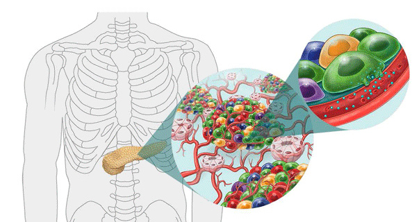
Type 1 diabetes (T1D) occurs when the insulin-producing beta cells in the islets of the pancreas are destroyed by the body’s own immune system. As a result, individuals with T1D must take daily insulin to control their blood glucose.
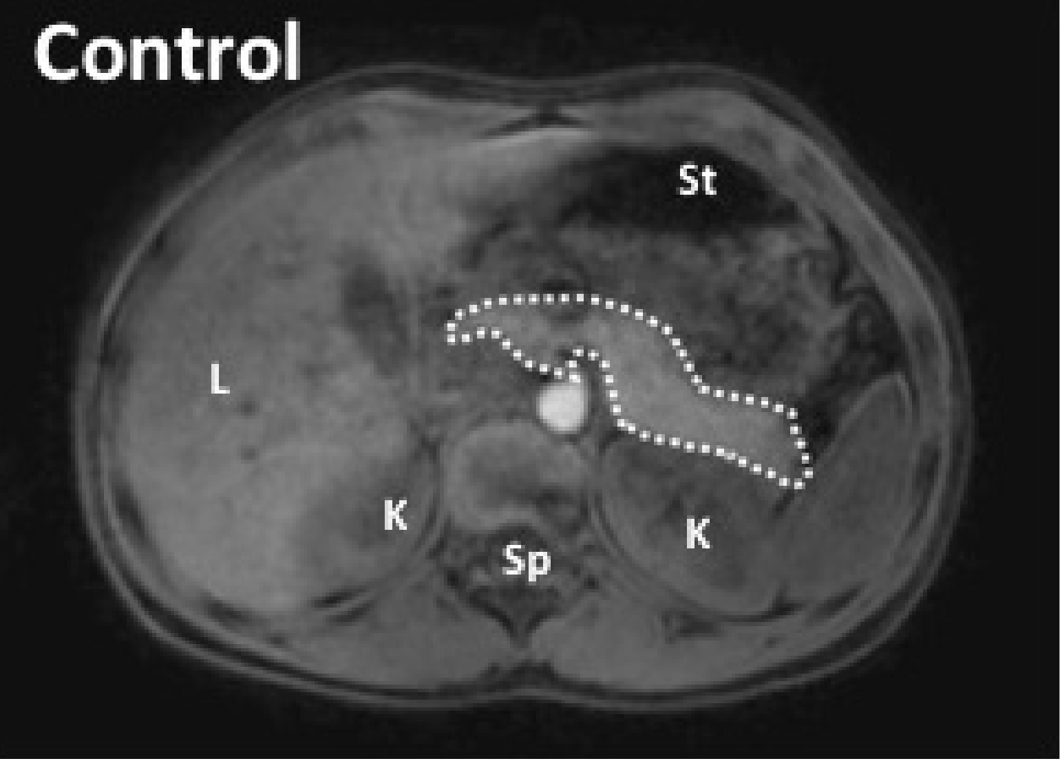
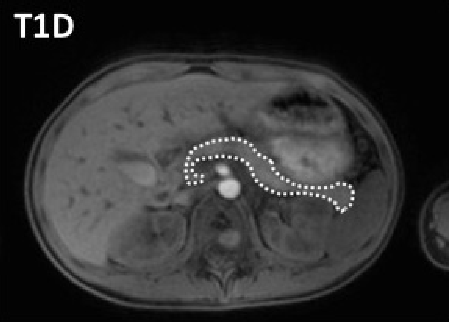
Magnetic resonance imaging (MRI) reveals that the size of the pancreas in people with T1D is reduced by approximately 30% compared to healthy controls. Only 1-2% of the mass of the pancreas is composed of islets, so the loss of pancreas volume cannot be solely explained by the destruction of beta cells.


MRI of abdomen of an individual with T1D and an individual without T1D (Control). The pancreas in both images is outlined with a dotted white line. On the left image, other abdominal organs are noted with a letter: L (liver), St (stomach), K (kidney), Sp (spine). Diabetes Care 42: 248, 2019


