Vanderbilt Dermatology Translational Research Clinic
Making skin measurements
for clinical impact
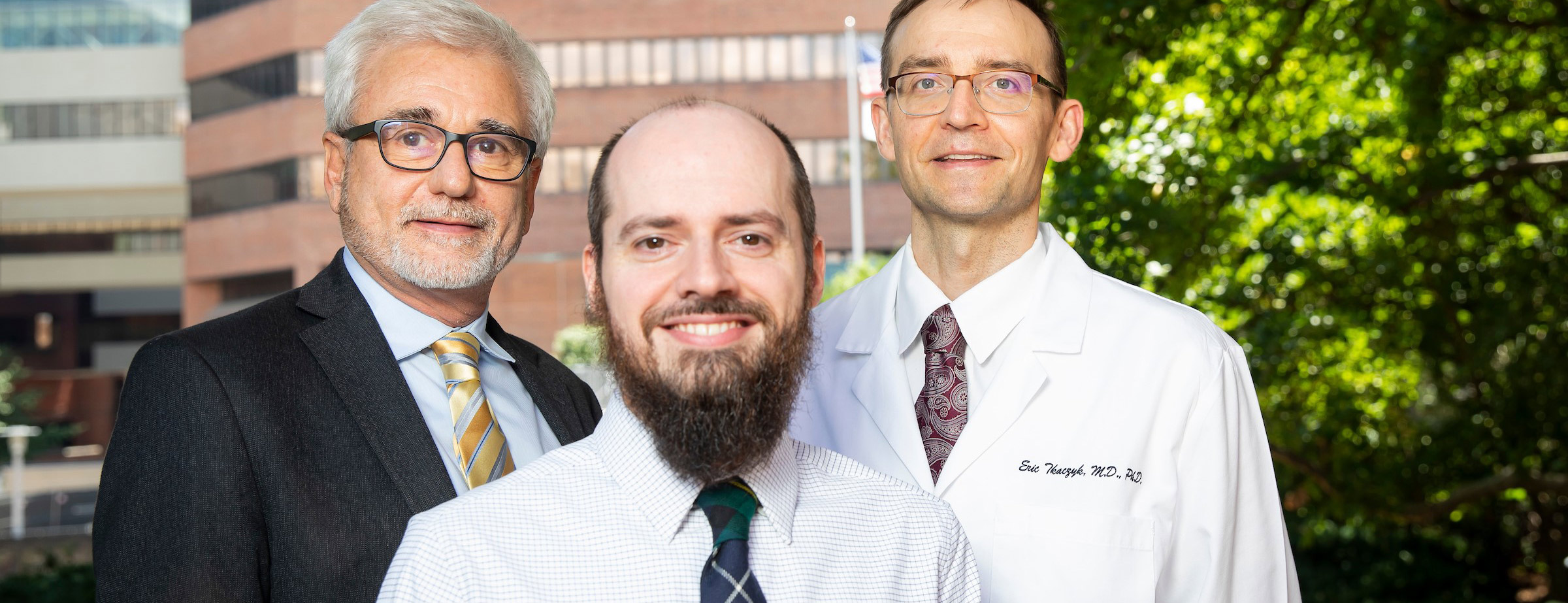
The central focus of the Vanderbilt Dermatology Translational Research Clinic (VDTRC), led by Dr. Eric Tkaczyk, is the development and application of technology for rigorous, clinically meaningful assessment of the skin, both for cutaneous as well as systemic diseases.
Full-Time Positions Available
Full-time positions are available immediately for those preparing for their next competitive stage of training (graduate and/or medical school, MD/PhD, dermatology residency, or faculty position).
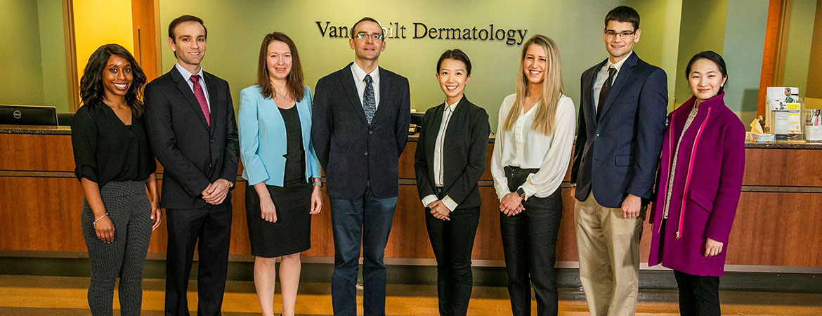
We are a multidisciplinary team of clinicians and engineers whose mission is to seamlessly integrate patient care, clinical investigation and fundamental technology research with applications in dermatology, oncology, hematology, and other specialties.
The VDTRC research program integrates development, application, and validation of skin assessment tools. Many clinical breakthroughs would be possible by properly harnessing technology for diagnosis and treatment monitoring.
As the skin has been used since ancient times as a window into health, dermatology stands poised as the model specialty to transform patient care with objective measurements.
Skin Photo Analysis by AI
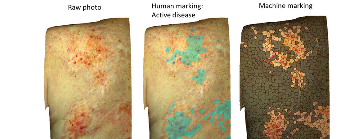
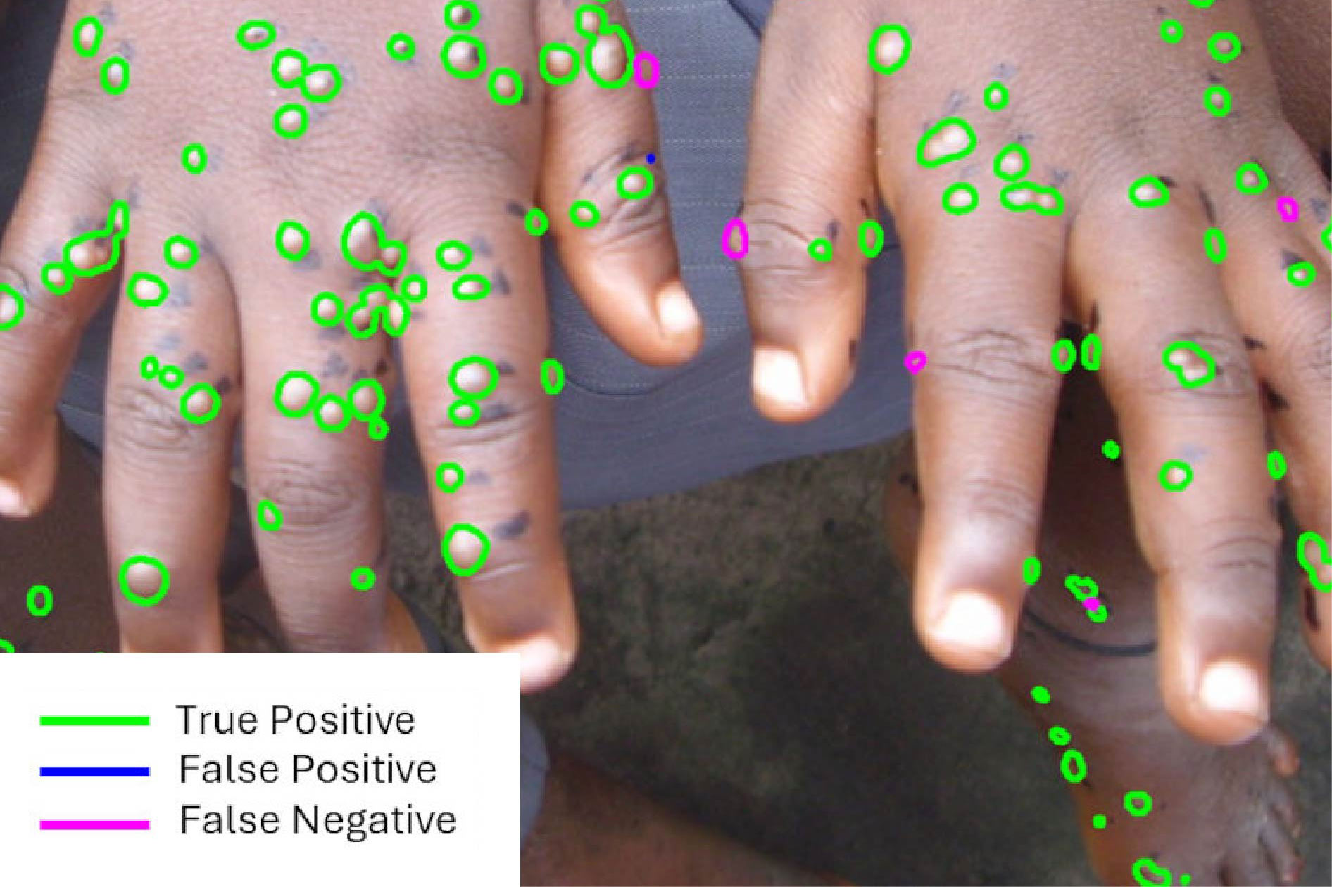
We are validating AI technology that was developed to measure skin erythema quickly and reproducibly using photographs. This is important because skin erythema is a trackable biomarker strongly associated with long-term mortality of chronic graft-versus-host disease (GVHD).
The clinic develops and supports the photography and skin scoring protocols for multicenter randomized controlled trials in Stevens-Johnson Syndrome and mpox (formerly monkeypox).
McNeil AJ, House DW, Mbala-Kingebeni P, Mbaya OT, Dodd LE, Cowen EW, Nussenblatt V, Bonnett T, Chen Z, Saknite I, Dawant BM, Tkaczyk ER. Counting monkeypox lesions in patient photographs: limits of agreement of manual counts and artificial intelligence. Journal of Investigative Dermatology. 2023 Feb 1;143(2):347-50. PMID: 36116509 PMCID: PMC9534148 DOI: 10.1016/j.jid.2022.08.044
Mechanical Measurement of Sclerosing Disease
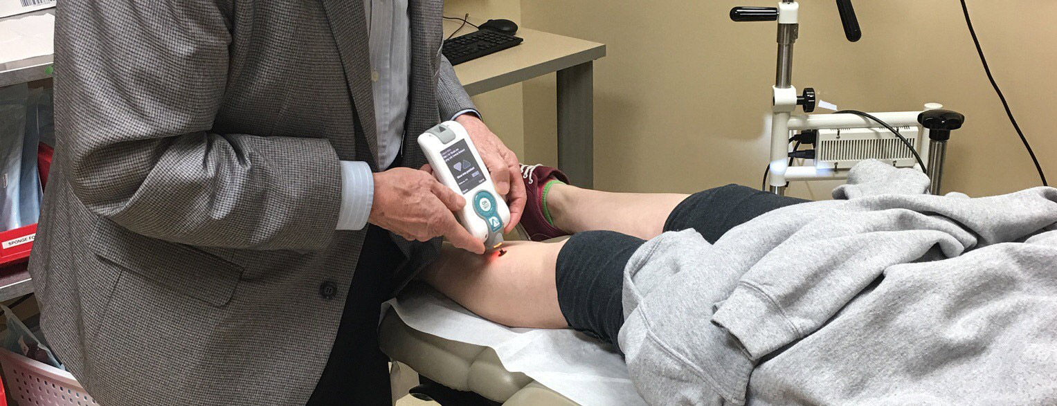
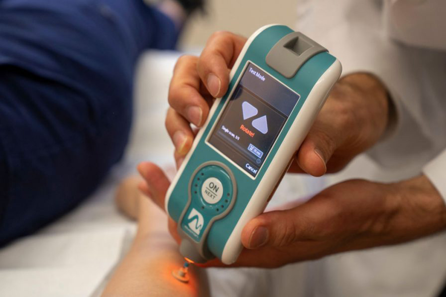
To address the unmet need to track the efficacy of antifibrotic therapy of sclerotic GVHD and systemic sclerosis, the VDTRC pioneered handheld devices to measure skin mechanical parameters.
The clinic’s experimental work includes microscopy, imaging from hyperspectral cameras to smartphones, ultrasound and investigations of biomechanics of fibrotic disease.
Patient Care
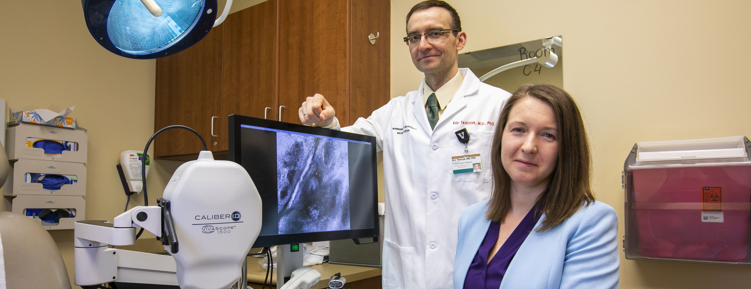
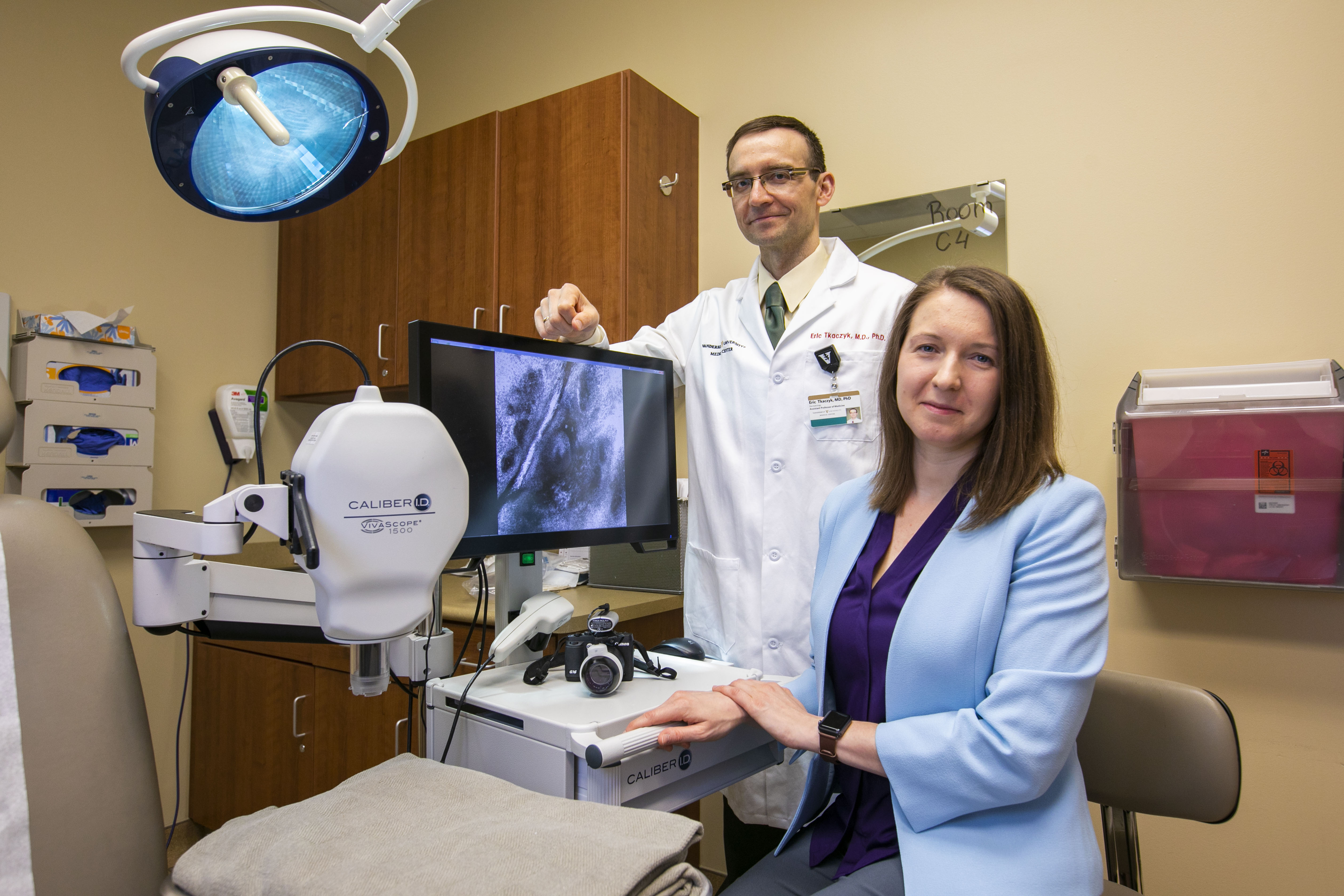
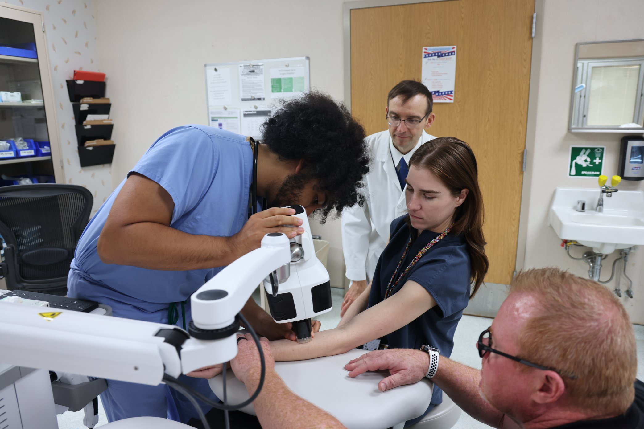
Within the Department of Dermatology at Vanderbilt University Medical Center, we offer noninvasive skin biopsies with reflectance confocal microscopy (RCM)
- Bladeless
- Painless
- Approved for reimbursement by Medicare and many other insurances
- “Cutting-edge technology without the cutting”

